Weird Case Files: A rare feline cancer

Welcome to our blog series, “Weird Case Files”, where our Associate IndeVets discuss their favorite + fascinating clinical cases.
One morning in late November of 2023, the practice manager at one of my regular clinics came to me with a question. A client was calling about Max, their very ill cat; they thought he might be dying and wanted to ease his passing. He had been seen a few months prior at the emergency room, according to the owner, and at that time was diagnosed with presumptive cancer. Our schedule was full for the day and technically he was not a current patient, as he had not been in for an exam for several years, but was there a chance we would be able to fit this cat in for euthanasia? I reviewed the schedule, checked in with my technicians, and affirmed that we could indeed accommodate the client’s request a bit later that morning.
Shortly before the cat arrived, I reviewed the ER report:
7/25/23
History
Max presents for an open wound between his legs. O states she noticed a lump between Max’s legs a month ago, and now Max has licked it so much it is an open wound. O said she noticed it was open on Thursday and put a cone on him. Still eating and drinking. No vomiting or diarrhea. History of herpes, no daily medications, no seizure history. NOT UTD [on vaccinations].
Vitals
Temp 101.4. HR 188. RR 36. MM pale pink, semi-moist, CRT 1-2 sec. Weight 6.3 kg, BCS 8/9
Physical Exam
Temperament: Friendly & Calm
Hydration: 5% dehydration; semimoist, tacky membranes and minimal loss of skin turgor
Eyes: No significant findings Ears: No significant findings Nose: No significant findings
Oral Cavity: No significant findings
Neck and Thyroid: No significant findings
Lymphatic: No peripheral lymphadenopathy
Cardiovascular: Normal heart rate, sinus rhythm, no murmurs. Strong and synchronous pulses bilaterally.
Respiratory: Normal respiratory rate and effort. Auscultation of lungs – No significant findings.
Abdominal: Soft and nonpainful. No palpable masses, organomegaly or fluid wave noted
Musculoskeletal: Ambulatory x 4 with no apparent pain, lameness or joint effusion. Normal muscle mass and tone. No neck or back pain appreciated.
Urogenital: Abnormal; Large firm irregular mass at prepuce, unable to exteriorize penis. Ulcerated ventral aspect with surrounding moist dermatitis
Mammary: No significant findings
Integumentary: Abnormal; Dermal mass at prepuce, ulcerated with moist dermatitis
Neurological: No abnormalities appreciated
Rectal: Not examined
Assessments
Preputial mass – r/o sarcoma vs other
Diagnostics – none
Plan
Discussed concern for mass, unable to appreciate if impacting the penis/urethra. Can consider supportive care pending follow up with surgeon (discussed considerations with boarded surgeon v. generalist, attempt at curative surgery v. debulking). O approves medications pending follow up for surgery. Discussed that if p appears to decline or is unable to urinate, compassionate euthanasia can be reasonably considered
- Convenia 8 mg/kg SQ
- Simbadol 0.12 mg/kg SQ
11/29/23
History
Presenting for euthanasia. Has not eaten in five days and has lost a significant amount of weight. Has a growth near his penis and another on his leg. Per ER doctor, preputial mass could cause difficulty urinating – owner thinks that is happening. Lays in litter box, but no urine has been produced. Is not drinking water, and is not interested when owner tries to encourage him to drink. Owner has noticed a significant decline in his quality of life over the last 1-2 weeks.
Owner also notes that one of Max’s littermates died earlier in the year from metastatic neoplasia.
Attitude / Appearance
BAR, mm pink, tacky, CRT<2s. Decreased skin turgor. Marked muscle wasting.
Weight: 4.27 kg; BCS 2/9
Behavioral/Fear, Anxiety, Stress Assessment: FAS 1/5. Very sweet cat.
Pain Score 2-3/4. Temperature not taken in keeping with Fear Free handling.
Objective
Oral Cavity/Teeth: Not performed due to FAS
Eyes/Orbit: OU – Mild blepharospasm and moderate mucoid discharge. Patchy cloudy corneal opacity OD. Fundic exam not performed.
Ears: AU – No aural inflammation or ceruminous debris; otoscopic exam not performed.
Throat: No appreciable thyroid slip. No cough elicited on tracheal palpation.
Cardiovascular: HR 180. No murmur or arrhythmia ausculted, femoral pulses strong and synchronous.
Respiratory: RR 30. Eupneic, normal bronchovesicular sounds diffusely. No nasal discharge.
Abdomen: Soft/non-painful. Cranial abdominal organomegaly noted. No appreciable fluid wave.
Musculoskeletal: Ambulatory x 4; No joint effusion noted; No long bone or joint pain evident. Diffuse muscle wasting.
Glands/Lymph Nodes: Mandibular, prescapular and popliteal lymph nodes palpated – diffuse mild peripheral lymphadenopathy noted.
Urogenital: 2.5 cm irregular firm preputial dermal mass present.
Rectal: Not performed.
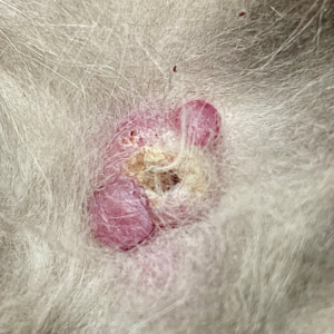
Integumentary: Healthy haircoat, decreased skin turgor. No appreciable ectoparasites. Multifocal dermal nodules in size ranging from 0.25 cm (on face) to 2.5 cm (prepuce). Nodules are scattered throughout dermis all over cat. Some are ulcerated, some scabbed.
Nervous System: Mentally appropriate. No ataxia. No neurologic deficits or abnormalities. Complete neurologic exam/assessment not performed today.
Assessment
10 yo MN DSH presenting for humane euthanasia
A1) Multifocal dermal nodules r/o mast cell tumor, histiocytic sarcoma, cutaneous lymphoma, mycobacterium, granulomatosis, other
A2) MM wasting / severe weight loss
A3) Inappetence
A4) Cranial abdominal organomegaly r/o infiltrative disease, reactive/hyperplastic splenic change, other
A5) Dehydration
A6) Ocular disease (rule-out list not detailed as pet presenting for euthanasia
Plan
Humane euthanasia
Although I was able to provide Max with a gentle passing and relief from his suffering, I did not have answers as to what had caused his illness. Clearly this was an aggressive, multifocal disease process, but what was it? Cancer was high on my rule-out list, given Max’s age, appearance, and status as an indoor cat (thereby making an infectious etiology less likely).
In evaluating him again post-mortem, I noted that in addition to the dermal masses all over his body, his paw pads all looked slightly puffy. They were reminiscent of “pillow foot”, or feline plasma cell pododermatitis, although not nearly as severe as the cases I had seen.
My curiosity was piqued – in over fifteen years as a veterinarian, I had never seen a case like this one. As a profession, we dislike going without answers. Even though any information I obtained would come too late for Max, there was always the possibility of seeing another case like this again some day.
Clinical pathology has always been a favorite subject of mine, and I carefully made a couple of slides from the dermal masses in the hopes that they would provide some clarity.
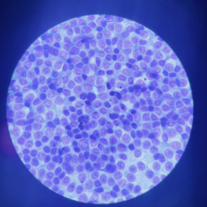
It was immediately apparent that Max had indeed been dealing with a neoplastic process.It appeared to be a round cell tumor, but beyond that, I was unable to be more specific. Cutaneous lymphoma came to mind, though this was not consistent with its usual appearance in cats. Although I saw no mast cell granules inside the cells, poorly-granulated MCT are notoriously aggressive, and would also fit the presentation.
I checked in with my fellow IndeVets, providing them with the history, photos, and cytology images. The general consensus was that my two rule-outs fit, but one of my colleagues (hat tip to Dr. Carla Germano) suggested a condition with which I was unfamiliar: FPH, or feline progressive histiocytosis. Brief research revealed that FPH did indeed fit the clinical picture. According to the UC Davis VetMed website:
- Feline Progressive Histiocytosis (FPH) is a disease of middle-aged to older cats, the age ranged from 7 to 17 years (Vet Pathol. 2006;43(5):646–655). The initial presentation of FPH may be a solitary skin nodule, although usually multiple papules, nodules or plaques develop, measuring up to 1.5 cm in diameter. The nodules are firm, non-pruritic and non-painful. The surface is often alopecic and may be ulcerated. The lesions are mostly located on the head, lower extremities or trunk. Occasionally, the lesions are limited to one extremity. The lesions may wax and wane but spontaneous regression does not occur. In FPH, a proportion of cats develop invasive, expansile masses in lymph nodes and internal organs including the lungs, kidneys, spleen, and liver.
In preparation for writing this article, I submitted Max’s case to a clinical pathology service in the hopes of obtaining further clarification. Their differential list matched mine: histiocytic disease (such as FPH), atypical cutaneous lymphoma, and poorly-granulated mast cell neoplasia. Determining which of the three we had been dealing with would have required histopathology, which was not performed. Further clarity would remain elusive, but I did have confirmation that we had been on the right track, diagnostically speaking.
Three potential diagnoses, none with a good prognosis. At the time he presented for euthanasia, it had been just over four months since Max’s ER visit, and roughly five months since the first mass had been noted at home. Given that cats are notorious for hiding medical problems from their owners, it was entirely possible that the initial tumor had been present for longer than that.
Unfortunately, Max’s story did not have a happy ending. I am grateful for what he enabled me to learn and will carry that knowledge through the rest of my career. As clinicians and caregivers, we owe it to our euthanasia patients to not only humanely end their suffering, but to use the insights they gift us when caring for future patients. Thank you, Max.