Eggs-traordinary Care: “Winnie” the Python’s Surgical Success


Meet Winifred Ssssanderson: Ball Python
When Winifred Ssssanderson walked into the hospital, she was in trouble. Well, “walked in” wasn’t exactly accurate. After all, this lady doesn’t have legs.
She was carried in a box. This beautiful lady was a ball python — a banana cinnamon genetic stripe to be exact — and she came to see me because she was in distress. Winnifred (Winnie) was a breeding snake who was cared for by a meticulous person who wanted to do right by her. Ball pythons typically start laying eggs between 4-6 years of age, and this gal was right on target. She should have laid her clutch three to four days before she saw us. The client knew something was wrong and came to us for help.
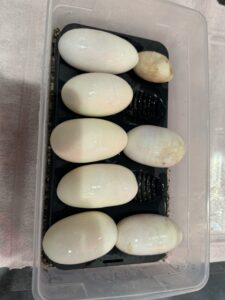
Upon opening the box, it was clear she was struggling. Winnie was stuffed to the brim with eggs. Their rounded edges strained against her sides – to the point of causing multifocal bruising. Rather than curling into the classic ball, Winnie shifted uncomfortably in an attempt to make as much space as she could for her own internal organs. She then lay as still as possible, as if she were walking on eggshells inside her own skin.
Diagnosing and Deciding on Winnie’s Treatment
As I spoke with the client, we discussed what we could do to help Winnie. Could we do surgery to save her? Were there baby snakes inside the eggs? Were they still alive?
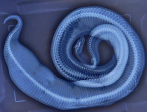
We began by obtaining x-rays which confirmed what we already knew. This kiddo was stuffed with shells and ready to pop. Their oval outlines peeked out at us from the dark screen, visible against the outline of her tubular frame. Snake eggs are usually poorly calcified, so their opacity in the caudal 1/3 of the patient can look like soft tissue. A classic egg-bound snake. Easy diagnosis . . . not necessarily an easy fix.
Veterinarians are smart people. We know how to problem solve. We can take what we already know and are comfortable with and apply it to a novel situation. We may not immediately know the answers to everything, but we sure know where to find them!
I had never spayed a snake before, but I had performed growth removals and laceration repairs on them. I informed the owner that the procedure itself in this species was new territory, but that I had cared for reptiles under general anesthesia previously, in addition to completing a VIN CE course on exotic animal surgery. The client was offered referral to a specialist and chose to have the surgery done with us that day. Take what you know and figure out the rest. Novel species spay? Challenge accepted!
Anesthetic Challenges and Surgical Strategy
Fortunately, snakes are extremely easy to intubate. The glottis lies in the front of the mouth, with a lovely hole just begging for an ET tube. As a ball python, Winnie – along with other snake and lizard species– had incomplete tracheal rings, so gently inflating the ET tube cuff was safe for her. If I had been anesthetizing a turtle or crocodile, that would have been a different story. Those groups have complete tracheal rings, so don’t puff that cuff! Most of the time, the trachea bifurcates just past the dorsal aspect of the heart. Palpating the heart can give you a pretty good landmark for how deep to place the ET tube. Watch the dead space! In general, snakes retain a functional right lung, so use caution to avoid placing the tube too deep and inadvertently entering the vestigial left lung. Diverse species have variable functional capacity and size of the “leftover” left lung.
Since Winnie didn’t have a functional muscular diaphragm (same as all the other reptiles, except crocodiles), she needed to have us breathe for her. When reptiles are conscious, the intercostal muscles provide most of the heavy lifting of respiration. For our anesthetized patient, this could be accomplished by a mechanical ventilator or a dedicated technician providing 4-8 breaths per minute. 10-15mmHg cm H20 should be peak airway pressure when ventilating a snake. Another important consideration for reptile anesthesia is keeping the patient warm. We already know how important this is for our mammalian patients; this goes double for ectotherms who rely on their environmental temperature to maintain homeostasis.
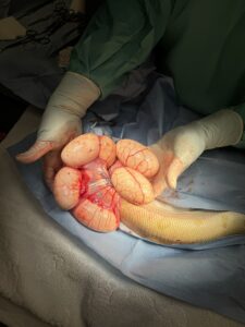
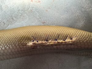
Once we had Winnie safely asleep and toasty warm, where do we make the incision? The key was to avoid the ventrum. We don’t want an iatrogenic wound trying to heal while it’s being constantly rubbed on. A lateral incision is also less likely to get contaminated during healing. There is also a ventral abdominal vein along the midline of snakes. I don’t know about you, but I prefer to avoid large blood vessels with a surgical approach if I can! I find it easiest to have snakes in lateral recumbency so I can get great visualization of the non-dependent side. Make a longitudinal incision between the first and second or second and third row of scales dorsal from the delineation between the ventral scutes and the lateral scales. Incise the skin between the scales, not through or on the scales themselves. This results in a “zigzag” pattern between the scales.
After making an incision, and gently maneuvering the reproductive tract to the opening, the eggs started to erupt from her body. Tracing the distended paired uterine horns, we discovered that two eggs had adhered together. There was no way they would have come out naturally! Fortunately, there was minimal bleeding, and Winnie maintained excellent vital signs throughout the procedure. We flushed her coeloemic cavity with warm sterile fluids, just in case there was any contamination.
But how to put her together again? Closing the body wall is similar to a closure in a mammal. Using absorbable monofilament in a simple continuous pattern tends to work well. But what about the skin layer? How does one close skin that is covered with scales? Everting suture pattern to the rescue! As this layer contracts during healing. Personally, I like horizontal mattresses. A vertical mattress or other everting pattern would work as well. Leave the sutures in for a good 6-8 weeks. They will frequently slough off with the next ecdysis. If they don’t, they can be removed.
Slow and Steady: Winnie’s Recovery from Anesthesia
Reptiles take a loooooooong time to do anything: get sick, show that they’re sick, respond to treatment, go under anesthesia, wake up from anesthesia, and recover from anesthesia. Winnifred was no exception.
It was a long recovery compared to a mammal, but perfectly on par for a python. Not only is their anatomy distinctive, but their physiology is unique as well. For our mammals, we leave the O2 on for a while in early recovery, just to top them off, right? Not so for reptiles! Respiration is stimulation by hypoxia — NOT hypercapnia as in a mammal — so keeping a snake on O2 longer actually translates to a sleepier snake with a slower recovery.
Winnie was so much more comfortable afterwards! She looked so content under her heat source. Her beautiful scaly self was able to move with ease, and she poked her head up frequently to see who else we were helping in the treatment room. A few minutes later, she had completely turned around and was facing the other way. This was a very good sign! Our patient was no longer in such pain that she had to stay in a “guarded” posture and was able to exhibit normal, fluid movement and chose to do so.
Even though removing Winnie’s eggs culminated in a decrease of one third of her presenting body weight, she doesn’t get to stuff herself with anything else for at least a week or two. The last thing you want is an entire rodent traveling through your patient and putting excess tension on the new surgical site by stretching it as it passes by, causing dehiscence from the inside out. If needed, tube feeding is an option.
I am grateful to have met and learned so much from helping Winnie. I’d call her story a surgical success!

References
Bennet, Avery, and Geoff Pye. “EXOT611-0522 Exotic Animal Surgery.” VIN CE. Exotic Animal Surgery, 5 May 2022.
Bennet, R. Avery, and Geoffrey W. Pye. Surgery of Exotic Animals. John Wiley & Sons, Inc, 2022.
Carpenter, James W. ad Craig A. Harms. Carpenter’s Exotic Animal Formulary. 6th ed., Elsevier, 2023.
De la Navarre, Byron. “Anesthesia of reptiles (Proceedings).” DVM 360. May 1, 2011.
https://www.dvm360.com/view/anesthesia-reptiles-proceedings
Divers, Stephen J., and Scott J. Stahl, editors. Mader’s Reptile and Amphibian Medicine and Surgery. 3rd ed., Saunders, 2019.
Grahm, Jennifer E, et al. Exotic Animal Emergency and Critical Care Medicine. John Wiley & Sons Inc, 2021.
Jepson, Lance. Exotic Animal Medicine. A Quick Reference Guide. 2nd ed. Elsevier, 2016
Mader, Douglas R. “Clinical anatomy of reptiles (Proceedings).” DVM 360. August 1, 2008. https://www.dvm360.com/view/clinical-anatomy-reptiles-proceedings-0.
Mayer, Jorg and Thomas Donnelly. Clinical Veterinary Advisor: Birds and Exotic Pets. Saunders. 2013.